A powerful diagnostic tool
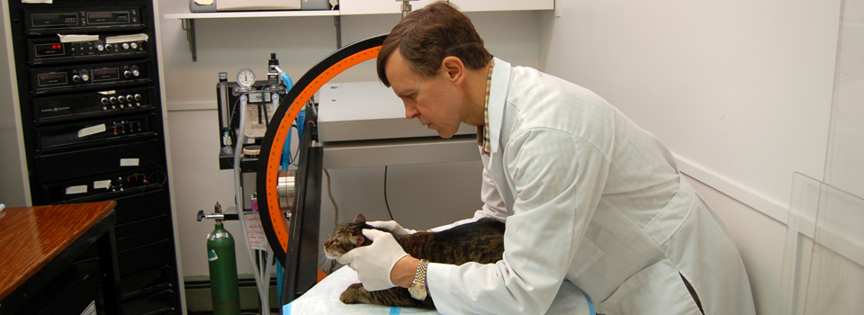
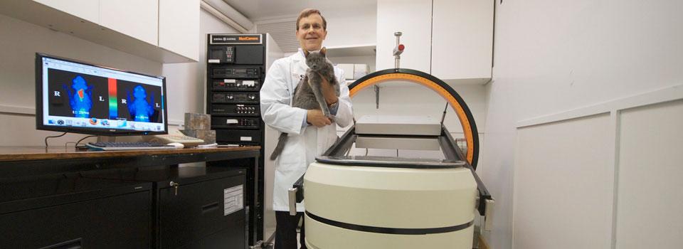
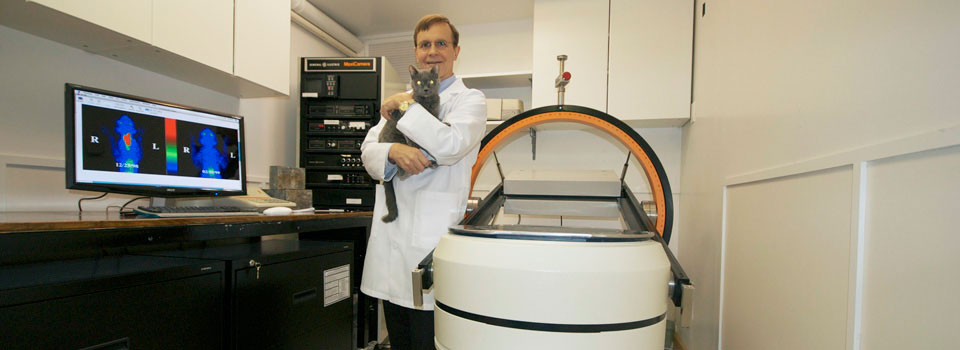
Nuclear Imaging for Animals is a specialized diagnostic imaging facility dedicated solely to scanning animals with nuclear medicine. Other than Cornell University, Nuclear Imaging for Animals is the only veterinary nuclear imaging facility in New York State. At the Animal Endocrine Clinic, we know that good diagnostic imaging goes a long way in making sure that your pet’s problems are diagnosed quickly and correctly.
What is nuclear imaging?
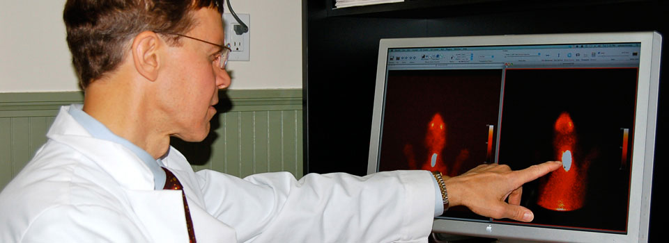
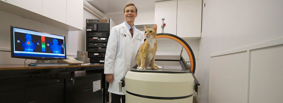
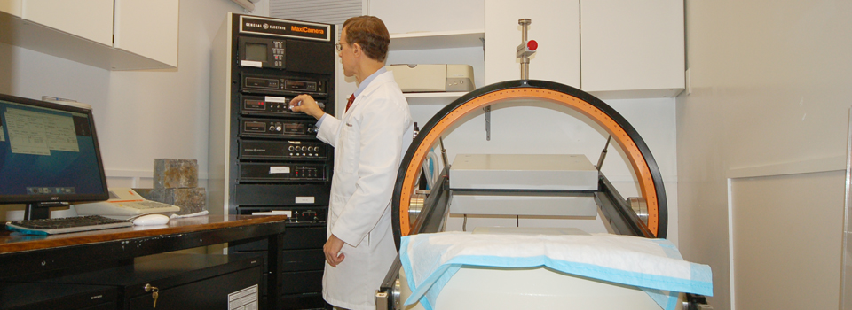
Nuclear imaging (scintigraphy) is a branch of radiology that provides crucial diagnostic information that can’t be derived from other imaging techniques such as radiographs (X-rays), CTs, MRIs, and ultrasounds. In nuclear imaging, we administer a small dose of a radioactive agent (radionuclide, radiotracer, or radiopharmaceutical) to the patient. These agents have no side effects; they are not dyes and are non-toxic. The radioactive agent is then taken up into specific tissues, where it emits gamma rays (electromagnetic waves similar to X-rays). A gamma camera detects these rays and uses them to form an image.
Why use nuclear imaging?
Nuclear imaging is a powerful diagnostic tool for two main reasons. First, it is organ and tissue specific. We use radiotracers specific for different tissues (i.e., a thyroid scan uses a different radiotracer than a bone scan). Only tissue of interest will take up the radiotracer, showing us only what we want to see. Second, nuclear imaging assesses both organ structure and function. Other imaging procedures can only assess anatomy (how organs look physically). This functional imaging is the strength of nuclear imaging; it can readily detect disease at an earlier stage than anatomical imaging procedures can.
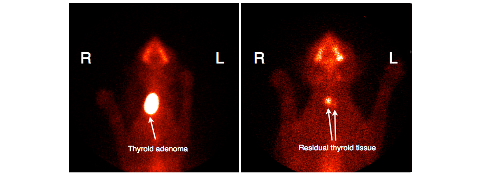
Because we deal primarily with animals that have endocrine disease, we mostly commonly do scintigraphy to image the thyroid gland. This is essential in the workup and staging of cats with hyperthyroidism, but is also very useful in evaluating dogs with thyroid disease (thyroid tumors or hypothyroidism).
Click here to read more about thyroid scintigraphy.